6 incredible images of the human brain built with the help of Google's AI
One of the ways artificial intelligence (AI) can be used is to help researchers make sense of huge amounts of data — including in science and medicine. We are members of a joint team of Google researchers and Harvard neuroscientists who have come together to build an unprecedented view into the human brain, which could help researchers understand neurological disorders and answer fundamental questions about how the brain works. By combining brain imaging with AI-based image processing and analysis, our teams have reconstructed nearly every cell and all of its connections within a small volume of human brain tissue about half the size of a grain of rice. Though it's of a small region of brain, this 3D mapping – which we’ve made freely available to the scientific community – nonetheless requires a monumental 1.4 petabytes (1.4 million gigabytes) to encode. Here’s what we found:
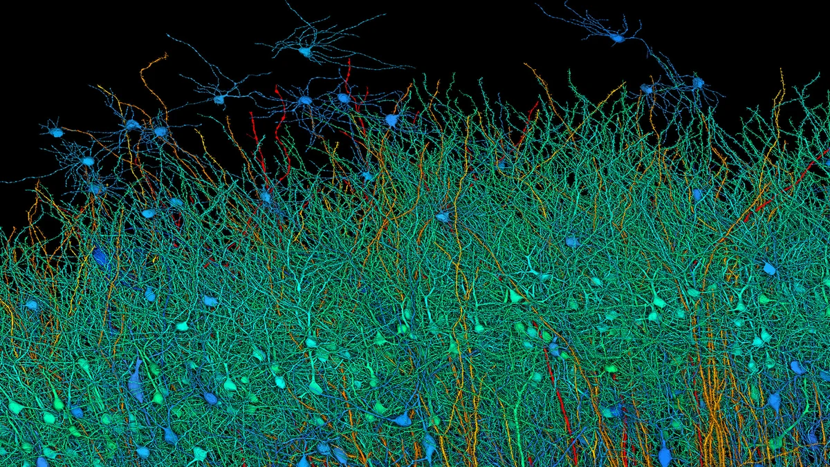
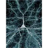
1. A view through six layers of the brain
Harvard researchers began by collecting thousands of extremely thin cross-sectional images from a donated brain sample. The small piece of healthy brain had to be removed during surgery on a woman with epilepsy to allow surgeons to reach the part they needed to operate on. A small piece of that otherwise discarded tissue was preserved as part of an IRB-approved study for later analysis.
The Google Research team developed advanced AI tools to construct an interactive 3D model of the brain tissue. The model underscores how complex the human brain is: describing just this small sample — one-millionth of the total human brain and about 3 mm long — requires more than a million Gigabytes of data: 1.4 Petabytes. This is the largest dataset ever made of human brain structure at this resolution.
Credit: Google Research & Lichtman Lab (Harvard University). Renderings by D. Berger (Harvard University)
The sample came from a part of the cortex (gray matter) called the anterior temporal lobe (see figure). The cortex has six layers, and by coloring the neurons according to their size and type, the layers are visible in this zoomed-out view of all of the neurons. The surface of the brain is at the top edge of the image.

Credit: Google
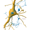
2. A dense and intricate map
The one cubic millimeter tissue sample contained about 50,000 cells and about 150 million synapses — the points of connection where signals cross from one neuron to another. Some neuron pairs had the surprising property of being connected to each other extremely strongly — through as many as 50 synapses. Researchers don’t know why they do this. This image shows a closeup of all of one type of neuron — the excitatory neuron — colored by size with red largest and blue smallest. The cells are about 15-30 micrometers across at their core.
Credit: Google Research & Lichtman Lab (Harvard University). Renderings by D. Berger (Harvard University)
3. A mirrored dance
An intriguing finding in the reconstruction was the existence of clusters of cells that tended to occur in mirror-image orientation to one another. This image shows a particularly symmetric pair.
Credit: Google Research & Lichtman Lab (Harvard University). Renderings by D. Berger (Harvard University)
4. Swimming in synapses
Neurons in the brain are intensely connected. This single neuron (white) has more than 5,000 axons (blue) arriving from other neurons to bring signals — and at least that many synapses where the signals cross from the axon to the receiving neuron. The synapses are shown in green.
Credit: Google Research & Lichtman Lab (Harvard University). Renderings by D. Berger (Harvard University)
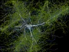
5. A curious finding: whorls of axons
A surprising finding of this research was the occurrence of “axon whorls.” Axons (blue) are the filamentous part of a nerve cell that carries a signal away from the cell. These looped piles of axon were rare in the sample, and in some cases they sat on the surface of another cell (yellow). Their function is unknown.
Credit: Google Research & Lichtman Lab (Harvard University). Renderings by D. Berger (Harvard University)
6. Serious networking
A single neuron (white) receives signals that determine whether or not the neuron fires. This image shows all of the axons that can tell it to fire (green) and all of those that can tell it not to (blue). Multiply this by the whole brain and that’s a lot of talking!
Credit: Google Research & Lichtman Lab (Harvard University). Renderings by D. Berger (Harvard University)
There is still a lot more to observe and understand from our reconstruction of this piece of human brain, and we hope other researchers will use the data to make additional discoveries. Scientists believe that by continuing research into the brain’s connections, they can one day understand things like how our memories form or what leads to neurological disorders and diseases like autism and Alzheimer’s.
Find out more about Google’s work on our Neural Mapping site, and learn more about this study at the Google Research Blog