How does oxygen move through brain? Scientists watch for the first time
Researchers have now, for the first time, developed a bioluminescence imaging technique that maps the movement of oxygen in the brain. The technique has been tested in the brains of mice.
")
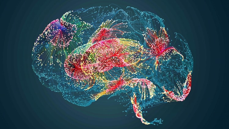
To map the movement of oxygen the team used luminescent proteins, chemical cousins of the bioluminescent proteins found in fireflies. (Photo: Getty)

New Delhi,UPDATED: Mar 29, 2024 13:03 IST
In Short
- The findings can help understand how oxygen travels in the brain
- It could open new doors to better understand and treat issues like hypoxia
- Existing oxygen monitoring techniques provide information about a small area of the brain
The human brain functions solely on energy generated by a form of metabolism that depends on oxygen, however, how it is distributed in the brain remained an enigma, until now.
Researchers have now, for the first time, developed a bioluminescence imaging technique that maps the movement of oxygen in the brain. The technique has been tested in the brains of mice.
advertisement
Mice share a significant portion of their genetic makeup with humans, making them valuable models for studying neurological diseases and disorders that also affect humans.
“This research demonstrates that we can monitor changes in oxygen concentration continuously and in a wide area of the brain. This provides us with a more detailed picture of what is occurring in the brain in real-time, allowing us to identify previously undetected areas of temporary hypoxia, which reflect changes in blood flow that can trigger neurological deficits” says Maiken Nedergaard, co-director of the Center for Translational Neuromedicine.
The findings can help understand how oxygen travels in the brain and could open new doors to better understand and treat issues like hypoxia, a condition characterised by a deficiency in the amount of oxygen reaching the tissues of the body.
To map the movement of oxygen the team used luminescent proteins, chemical cousins of the bioluminescent proteins found in fireflies.
While existing oxygen monitoring techniques provide information about a small area of the brain, the researchers observed, in real-time, the entire cortex of the mice. The intensity of the bioluminescence corresponded with the concentration of oxygen, which the researchers demonstrated by changing the amount of oxygen in the air the animals were breathing.
The team tried to understand what happens when small parts of the brain are denied oxygen for brief periods.
While monitoring the mice, the researchers observed that specific tiny areas of the brain would intermittently go dark, sometimes for several seconds, meaning that the oxygen supply was cut off. These areas, which the researchers named “hypoxic pockets,” were more prevalent in the brains of mice during a resting state, compared to when the animals were active.
“The door is open to study a range of diseases associated with hypoxia in the brain, including Alzheimer’s, vascular dementia, and long Covid, and how a sedentary lifestyle, ageing, hypertension, and other factors contribute to these diseases,” says Maiken Nedergaard.
The study published in the journal Science states that systematically exploring the response to various experimental conditions indicated that physical activity such as running reduces the occurrence of hypoxic regions.